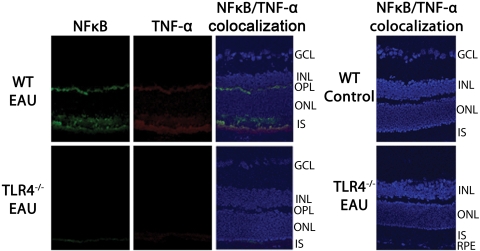
Figure 4.
Immunofluorescence localization of NFκB and TNF-α in the retinas of mice with EAU day 7 after immunization. Tissues were labeled using polyclonal anti-NFκB and monoclonal antibody against TNF-α (primary antibodies) along with Cy-2–conjugated donkey anti–rabbit IgG and Texas Red dye–conjugated donkey anti–mouse IgG, respectively (secondary antibodies). Nonimmunized WT and nonimmunized TLR4−/− mice showed no NFκB or TNF-α staining in the retina. During early EAU, however, WT mice showed NFκB and TNF-α staining in the inner segments of the photoreceptors and the outer plexiform layers, whereas such staining was absent in TLR4−/− mice.

