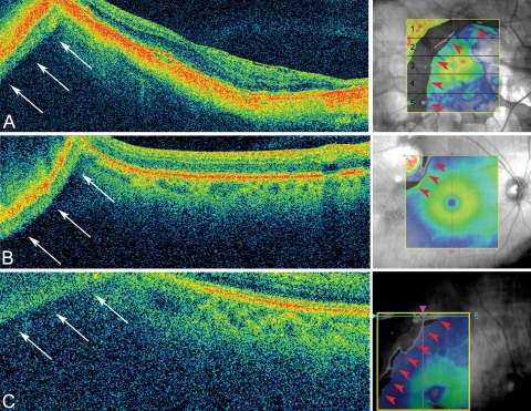
Figure 2.
Causes of mirror artifacts (all scans acquired using Cirrus macular cube 512 × 128. (A) A mirror artifact generated in a patient with high myopia is seen at the top left corner of the scan (arrows, left). A crescent-shaped defect is observed on topographic map due to the segmentation breakdown caused by the mirror artifact. The defect occupies five of five of the regions on the macular map (red arrowheads, right). This scan correlates to the light blue line on the topographic map (raster 64). (B) A mirror artifact caused by the thickening or elevation of the retina. Top left (arrows): the scan has flipped onto itself due to the presence of a superotemporal choroidal nevi. A segmentation defect is seen at the top left (red arrowheads, right). The scan (raster 1) correlates to the light blue line on the topographic map. (C) This patient was only slightly myopic (−0.63 D), but visual acuity was poor (20/200). In this case, the cause of the mirror artifact was due to retinal disease (neovascular AMD), which caused poor visual acuity and led to eccentric fixation. A segmentation defect was seen at the top left of the macular map (red arrowheads, right). The scan (raster 5) correlates to the light blue line on the topographic map.

