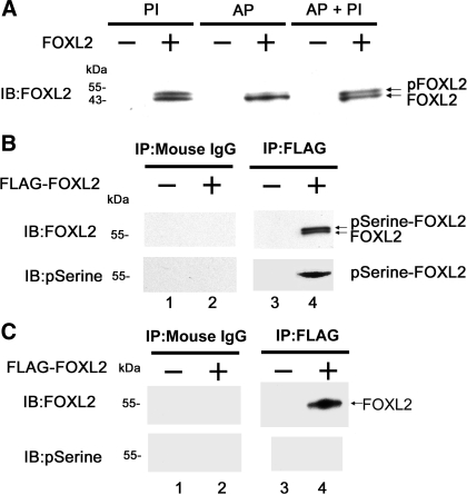
Fig. 2.
FOXL2 is phosphorylated in vitro. A: mammalian CHO cells, which do not express endogenous FOXL2, were transfected with the pcDNA3-FOXL2 expression construct (+) or with an empty pcDNA3 expression vector (−). Twenty-four hours after transfection, the cells were lysed and treated with phosphatase inhibitor alone (PI), alkaline phosphatase alone (AP), or AP and PI (AP + PI). The proteins were then analyzed by immunoblotting (IB). Two FOXL2 bands are seen in lysates treated with phosphatase inhibitor (PI), the lower of which corresponds in size to FOXL2 (41). The upper band was eliminated in lysates treated with AP but was preserved in lysates treated with AP + PI. B: CHO cells were transfected with pFLAG-FOXL2 expression construct (+) or an empty expression vector (−). Twenty-four hours after transfection, the cells were lysed and the resulting proteins immunoprecipitated with FLAG or mouse IgG (control) antibodies and analyzed by IB with FOXL2 and phosphoserine antibodies. Two FOXL2 bands were visualized using the FOXL2 antibody in the FLAG immunoprecipitates but not in the mouse IgG precipitates, the larger of which was also detected by the phosphoserine antibody (pSerine-FOXL2). C: lysates treated with AP in A were immunoblotted with the FOXL2 and phosphoserine antibodies. A single FOXL2 band was seen in the FLAG immunoprecipitates but not in the mouse IgG precipitates. No bands were detected using the phosphoserine antibody.

