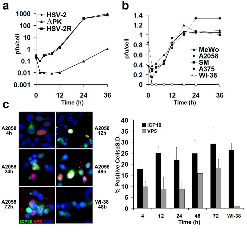
Figure 1. ΔPK is a growth-restricted replication competent oncolytic virus.
(a) Vero cells were infected with HSV-2, ΔPK, or HSV-2(R) (moi = 0.5) in serum-free medium and virus titers were determined by plaque assay. Results are expressed as mean pfu/cell (burst size). (b) A2058, MeWo, SM, A375 and Wi-38 cells were infected with ΔPK and examined for virus growth as in (a). Similar growth patterns were seen in melanoma cultures LM, SK-MEL-2, LN, OV and BUL. ΔPK did not grow in WI-38 cells and in normal melanocytes, but HSV-2 and HSV-2(R) replicated equally well in all the cultures. (c) ΔPK infected A2058 and WI-38, cells were stained with Alexafluor-488 labeled ICP10 and Alexafluor-594 labeled VP5 antibodies in double immunofluorescence. As described in Materials and Methods, ICP10 antibody recognizes both the wild type protein and the PK-deleted ICP10 protein, p95. Cells were counted in 3 randomly selected fields (≥ 250 cells) and the % staining cells calculated relative to total cells identified by DAPI staining. Quantitative results are shown for A2058 cell at 4-72 hrs p.i., and for WI-38 cells at 48hrs p.i. Similar results were obtained for the partially purified virus.

