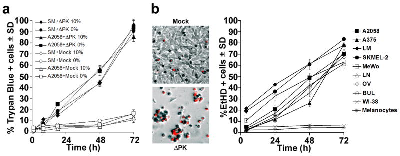
Figure 2. ΔPK-mediated melanoma oncolysis includes a robust PCD bystander component.
(a) A2058 and SM melanoma cultures infected with ΔPK (moi = 0.5) or mock infected with adsorption medium were cultured in medium without (0%) or with (10%) FBS and cells were stained with Trypan Blue at various times p.i. Four independent haemacytometer counts were performed and % staining cells was calculated. Results from 3 replicate experiments are expressed as mean % staining cells. (b) Melanoma, primary normal melanocytes and normal fibroblasts (WI-38) infected and cultured in serum-free medium were stained with EtHD. Cells were counted in 3 randomly selected fields (≥ 250 cells) and the % staining cells calculated as in (a). The image panels are ΔPK infected A2058 cells at 72h p.i. and are representative of all the melanoma cultures. Similar results were obtained for the partially purified virus.

