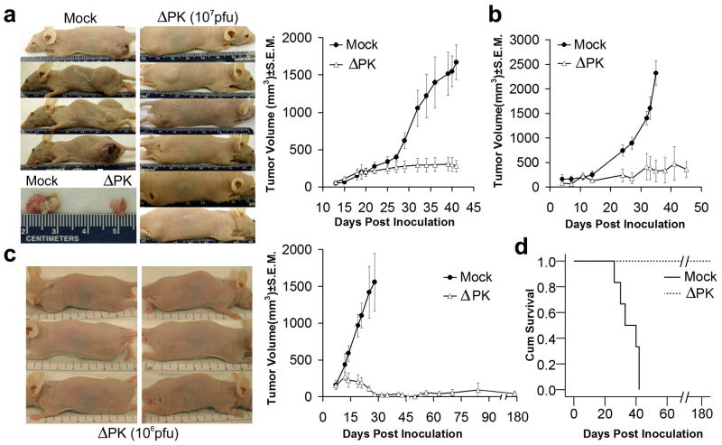
Figure 5. ΔPK inhibits the growth of melanoma xenografts.
(a) A2058 melanoma cells (107) were implanted subcutaneously into both flanks of Balb/c nude mice and given intra-tumoral (i.t.) injections (100μl) of ΔPK (n=12; 107 pfu) or growth medium (n=6; mock) beginning on day 14, when the tumors were palpable (approximately 200mm3). A total of 4 injections were given once weekly and tumor volume was calculated as described in Materials and Methods. The difference between mock and ΔPK treated animals became statistically significant on day 32 (p<0.001 by 2-way ANOVA) and remained significant to the end of the study. Representative animals and tumor tissues were photographed at day 42. (b) A375 xenografts were established as in (a) and given 4 i.t. injections of ΔPK (n=6; 106 pfu) or growth medium (n=6) at weekly intervals beginning on day 7, when the tumors were palpable. The difference between mock and ΔPK treatment became statistically significant at day 23 and remained significant by the end of the study (p<0.001 by 2-way ANOVA). (c) LM melanoma cells (107) were implanted subcutaneously into both flanks of Balb/c nude mice and given 4 i.t. injections of ΔPK (n=6; 106 pfu) or growth medium (n=6; mock) at weekly intervals beginning on day 7, when the tumors were palpable. Tumor volume in 4 animals was monitored for 5 months after the last ΔPK injection. The difference between mock and ΔPK treatment became statistically significant on day 14 (p<0.001 by 2-way ANOVA) and remained significant to the end of the study. Three ΔPK-treated mice showing complete tumor eradication were photographed at day 35. (d) Kaplan-Meier survival analysis in animals given LM xenografts with the terminal event set at 1.5cm diameter of growth in any one direction. ΔPK is significantly different from mock (p<0.001) by Log Rank (Mantel-Cox) analysis.

