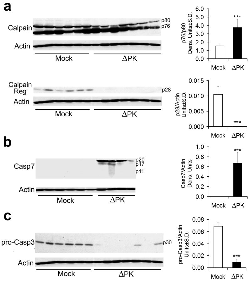
Figure 6. Calpain and caspases-7 and-3 are activated in ΔPK-treated xenografts.
A2058 xenograft tissues mock treated or treated with ΔPK as in Fig. 5a were collected 7 days after the last ΔPK injection and extracts were immunoblotted with antibodies to calpain (a) stripped and sequentially re-blotted with antibodies to activated caspase-7 (b), pro-caspase-3 (c) and actin. Each lane represents a different tumor. Representatives of three replicate experiments are shown for each antibody. Data were quantified by densitometric scanning as described in Materials and Methods, and results are expressed as densitometric units (*** p<0.001 vs. Mock).

