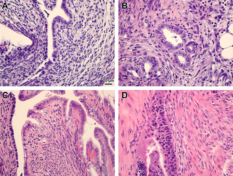
Figure 2. Histology of mouse endometriotic lesion and eutopic uterus.
Mice underwent surgical-induction of endometriosis and eutopic uteri (A, C) and endometriotic lesions (B, D) were excised 3 (A, B) or 29 (C, D) days later. Representative hematoxylin and eosin stained sections at 400× magnification are shown.

