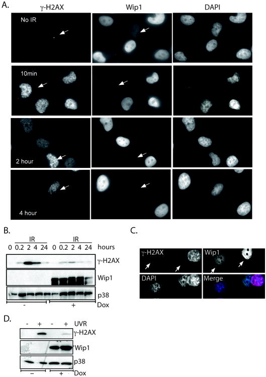
Figure 2. Wip1 did not block phosphorylation of H2AX after stress.
A) Immunocytochemistry of H1299 cells induced to express Wip1 either sham irradiated (“No IR”) or harvested at the indicated time points after IR exposure showed reduced γ̃H2AX foci at later time points. Arrows designate cells that do not express Wip1. B) Immunoblot analysis of the cell lysates from A) showed decreased IR-induced γ̃H2AX levels at later time points in H1299 cells expressing Wip1 (“+dox”) compared to the control (“-dox”). C) Immunocytochemistry showed reduced γ̃H2AX foci in Wip1 expressing H1299 cells compared to control cells (white arrows) after UVR exposure. (Wip1, green; γ̃H2AX, red; DAPI, blue) D) Immunoblot analysis of the cell lysates from C) showed similar results. The p38 immunoblot is used as a loading control.

