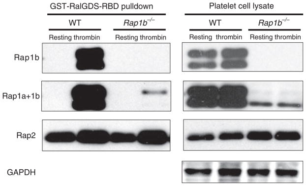
Fig. 3.
Thrombin stimulates Rap1a and Rap2 activation in Rap1b−/− platelets. Thrombin (1 U mL−1) or buffer was added to 6–8 × 10⁸ platelets mL−1 in modified Tyrode’s buffer for 3 min. Platelets were lysed, and equal amounts of lysates were used in a pulldown assay (left panels) followed by non-reduced sodium dodecylsulfate polyacrylamide gel electrophoresis and western blotting with Rap1b, Rap1a+1b (which recognizes both Rap1a and Rap1b) or Rap2 antibodies. Note that the Rap1b antibody does not crossreact with Rap1a in lysates from Rap1b−/− mice (Rap1b panels). Total cell lysates were used as loading controls (right panels). Data are representative of three independent experiments. GST, glutathione-S-transferase; RalGDS, Ral guanine nucleotide dissociation stimulator; RBD, Ras binding domain; GAPDH, glyceraldehyde-3-phosphate dehydrogenase; WT, wild-type.

