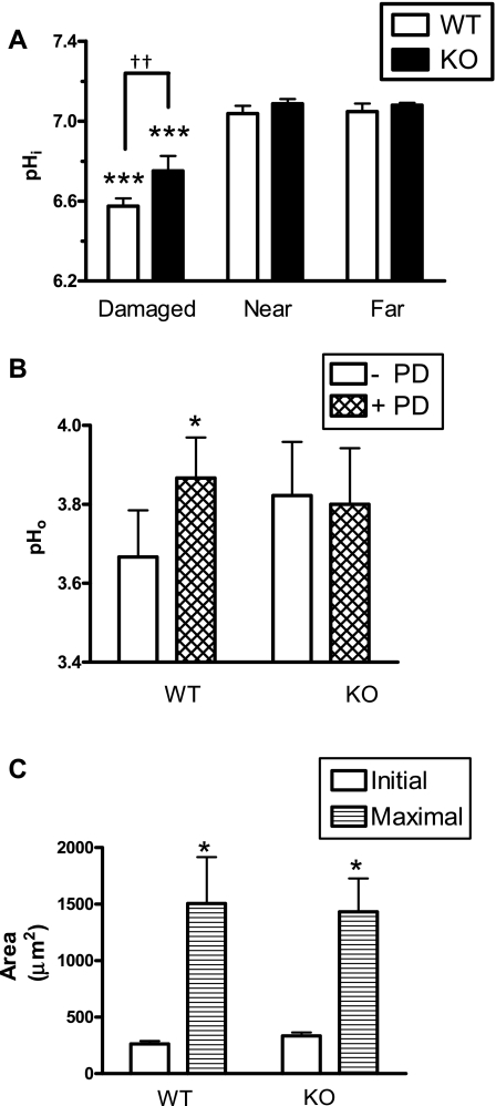
Fig. 8.
Intracellular pH, surface pH, and damage progression measured in solute-like carrier 26A9 (SLC26A9) wild-type (WT) and knockout (KO) animals. A: gastric epithelium of SLC26A9 WT (n = 4) or KO (n = 3) mice was loaded with SNARF-5F-AM, and pHi was measured before and after photodamage in damaged, near, and far cells in the same microscopic field. B: the gastric mucosa of WT (n = 9) or KO (n = 9) mice was superfused with pH 3 perfusate, and surface pH (pHo) was measured before and after photodamage. C: damage sizes were measured as those cells lacking NAD(P)H but retaining confocal reflectance. Initial size was measured immediately after photodamage, and maximal damage was evaluated over the first 10 min after photodamage. Means ± SE, *P < 0.05 vs. initial size; ***P < 0.001 vs. near cells; ††P < 0.01.

