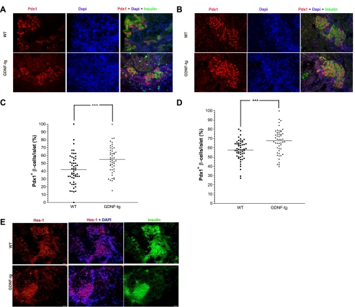
Fig. 5.
Pancreata from E18 and postnatal day 1 GDNF-tg mice have more Pdx1+ β-cells. A: pancreata from E18 GDNF-tg and WT mouse embryos immunofluorescently stained for Pdx1 (red) and insulin (green) with DAPI nuclear staining (blue). B: pancreata from postnatal day 1 GDNF-tg and WT littermates immunofluorescently stained for Pdx1 (red) and insulin (green) with DAPI nuclear staining (blue). Scale bar: 20 μm. C: score of Pdx1+ β-cells in islets from E18 GDNF-tg mouse embryos and WT littermates. Each point represents an islet. Horizontal lines show group means. ***P < 0.001, N = 3 WT and 6 GDNF-tg embryos. D: score of Pdx1+ β-cells in islets from postnatal day 1 GDNF-tg mice and WT littermates. Each point represents an islet. Horizontal lines show group means. ***P < 0.001, N = 3 WT and 5 GDNF-tg mice. E: pancreata from E18 GDNF-tg and WT mouse embryos immunofluorescently stained for Hes-1 (red) and insulin (green) with DAPI nuclear staining (blue). Scale bar: 20 μm.

