Fig. 5.
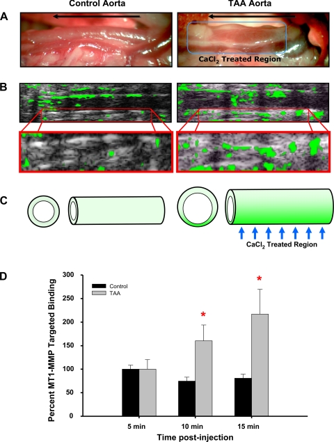
MT1-MMP-targeted microbubble contrast imaging. MT1-MMP antibody-conjugated microbubble contrast agent was injected into the tail vein and used to identify elevated MT1-MMP protein levels in the descending thoracic aorta of control and TAA-induced (8 wk) mice. A: intraoperative images of the descending thoracic aorta at terminal surgery showing increased aortic diameter at 8 wk post-TAA induction (right) versus a control animal (left). The direction of blood flow is shown by the arrow in each image, and the CaCl2 treatment region is shown on the right. B: high-resolution ultrasound imaging (equivalent region as shown in A) after the injection of MT1-MMP antibody-conjugated microbubble contrast agent (top). The green overlay indicates areas of enhanced MT1-MMP-targeted contrast binding. Specific binding was quantitated within a region of interest equivalent to the CaCl2-treated region in both control and TAA-induced animals. The bottom images show enlargement of the quantitated region of interest at 15 min after the contrast injection. C: pictogram summarizing the study results showing enhanced aortic dilatation accompanied by increased MT1-MMP-targeted microbubble contrast binding within the CaCl2-treated region. D: quantitative results of MT1-MMP-targeted microbubble contrast imaging demonstrating elevated binding at 10 and 15 min after the contrast injection. *P < 0.05 vs. 5 min. Representative images are shown.

