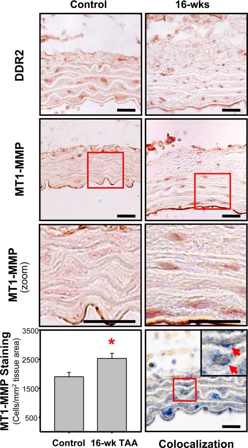
Fig. 6.
Immunohistochemical localization of discoidin domain receptor 2 (DDR2) and MT1-MMP. Immunostaining of 3-μm aortic tissue sections from control and TAA-induced mice (16 wk post-TAA induction) demonstrated the presence of DDR2-positive cells (top) and MT1-MMP-positive cells (middle top and middle bottom) within the aortic wall. Bottom left: quantitation of MT1-MMP immunostaining revealed an increased number MT1-MMP-positive cells in the 16-wk TAA-induced sections (33.3% increase vs. control, *P < 0.05). Bottom right: dual sequential immunostaining demonstrated the colocalization of MT1-MMP (blue) with DDR2 (brown) in aortic fibroblasts. Representative images are shown. Bars = 20 μm.

