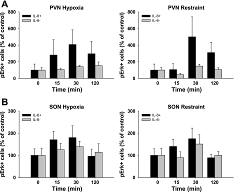
Fig. 8.
Effect of acute stress on ERK phosphorylation in IL-6- and IL-6+ neurons. Data are presented as a percentage of control (0 min) for the response to hypoxia (left) and restraint (right) in the PVN (A) and SON (B). There was a significant main effect of time on pERK induction in IL-6+ neurons that did not occur in IL-6- neurons (n = 6).

