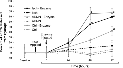
Fig. 4.
Hydrogels loaded with fluorescently tagged eEPC injected into mouse ears show the progression of cell mobilization over time. Graphical image shows the percent increase in release of fluorescently tagged eEPC from hydrogels injected into the ears of 3 groups of mice, adriamycin-treated (ADMN), unilateral renal ischemia (Isch), and control (Ctrl). Digestive enzyme injection (hyaluronidase and collagenase) into HA hydrogels decreased the number of visible cells in the ear, while eEPC not subject to dissolution by enzyme were prone to remain in hydrogels. *P < 0.05 vs. 0 and 24 h; #P < 0.005 vs. 0 h; n = 4.

