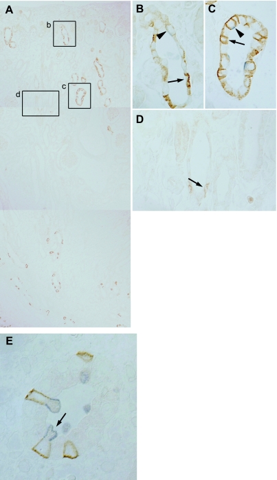
Fig. 5.
Double-immunolabeling, Rhbg and pendrin, at P4. A: composite micrograph showing entire kidney, from cortex to medullary tip. Rhbg immunolabel is present in a subset of cells in the cortex and the medullary tip, and is absent from intermediate regions. B: high-power micrograph of inset b in A showing convoluted epithelial tubule structure from the outer cortex. Cells with intense basolateral Rhbg immunolabel (arrow) do not express detectable pendrin and are identified as A-type intercalated cells. Pendrin-positive cells (arrowhead) express very low levels of basolateral Rhbg and are identified as B-type intercalated cells. C: high-power micrograph of inset c in A showing CNT closer to the corticomedullary junction that demonstrates that both A-type intercalated cells, with intense basolateral Rhbg but not apical pendrin (arrow) immunolabel, and non-A, non-B cell cells, with apical pendrin and basolateral Rhbg immunolabel (arrowhead), are present. D: high-power micrograph of inset d in A showing collecting duct from the deep cortex showing that only rare A-type intercalated cells, with basolateral Rhbg and without detectable pendrin immunolabel (arrow), are present. Rhbg immunolabel intensity is low, consistent with an immature phenotype of intercalated cells at this time point. E: micrograph from the deep medulla. Cells with intense basolateral Rhbg immunolabel (brown) express apical H+-ATPase immunolabel (blue), identifying these cells as A-type intercalated cells (arrow).

