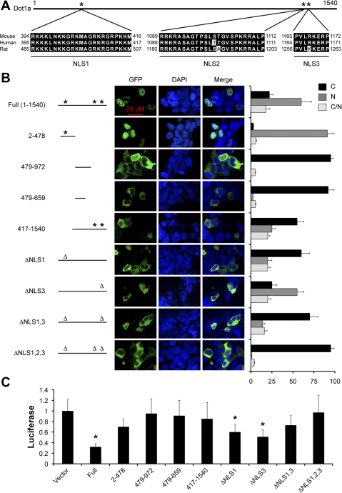
Fig. 1.
Dot1a has 3 potential nuclear localization signals (NLSs). The potential NLSs of Dot1 proteins from mouse, human, and rat (GenBank nos.AY196089, AF50950, and XP_343160, respectively) were identified using PSOFTII Prediction and aligned by MacVector software, with amino acid positions as indicated (A). Wild-type (WT) and various Dot1a mutants were transiently expressed as green fluorescent protein (GFP) fusions in 293T cells and analyzed by epifluorescence and confocal microscopy. DAPI, 4′,6-diamidino-2-phenylindole. Cells were categorized as cytoplasmic [C], nuclear [N], or both [C/N], depending on the predominant location of the fusion proteins. The presence (*) or absence (Δ) of a NLS was indicated. The graphed value (percentage) is the number of cells of each localization type divided by the total number of cells examined. At least 240 transfected cells/transfection were examined from 5 independent experiments (n = 5). M1 cells were transiently transfected with an α-subunit of the epithelial Na channel (αENaC) promoter-luciferase reporter along with a GFP vector (vector) or one of the GFP-Dot1a constructs as in B, followed by luciferase reporter assays (C); n = 3. *P < 0.05 vs. vector.

