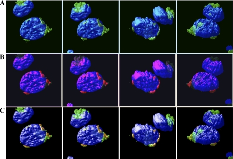
Fig. 5.
Cdk2-F80GmCherry localization with Golgi. TKPTS cells were transduced with Cdk2-F80GmCherry expression adenovirus. After 18–24 h, cells were fixed and stained for Golgi using anti-GM130 antibody and anti-mouse IgG-Alexa 488 secondary antibody. Cells were then stained with DAPI. Cells were scanned with LSM510 confocal fluorescence microscopy with a ×63 oil objective. To obtain the 3-dimensional (3D) images, Z-stack images taken by the confocal (0.5-μm slices) were used in Huygens Professional software (Scientific Volume Imaging). Localization of Golgi (green; A), Cdk2-F80GmCherry (red; B), and nuclei (blue). Colocalizations of Golgi and Cdk2 appear yellow/orange (C).

