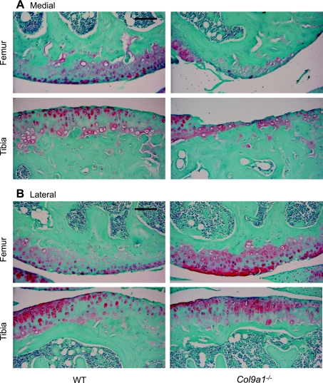
Fig. 1.
Sagittal sections of the femoral and tibial articular surfaces from 17-mo-old wild-type (WT) and Col9a1−/− mice. Representative sections are shown for the medial (A) and lateral (B) compartments of the knee. A: in the medial compartment, Col9a1−/− mice have significant cartilage loss, whereas, only mild superficial fibrilation is observed in WT mice. B: in the lateral compartment, both Col9a1−/− and WT mice show minor cartilage structural changes and loss of Safranin-O staining. Sections were stained with hematoxylin, fast green, and Safranin-O. Images are ×200 magnification (scale bars = 100 μm).

