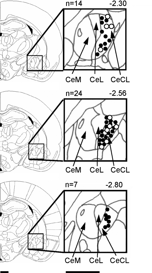
Fig. 1.
Histologically verified recording sites of 45 neurons in the laterocapsular division of the central nucleus of the amygdala (CeLC) recorded in 45 rats. Filled circles refer to CeLC neurons that were tested in pharmacologic studies (n = 34). Open circles show additional neurons (n = 11) that were characterized without pharmacologic tests. Boundaries of different amygdala nuclei are easily identified under the microscope. Diagrams (adapted from Paxinos and Watson 1998) show coronal brain sections at different levels posterior to bregma (−2.30 to −2.80). Next to each diagram are shown in detail the central nucleus and its medial (CeM), lateral (CeL), and laterocapsular (CeLC) subdivisions. Calibration bars are 1 mm.

