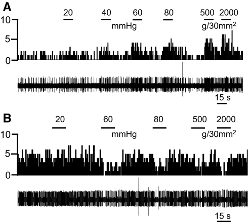
Fig. 2.
Visceronociceptive CeLC neurons. Background and evoked activity recorded in 2 individual CeLC neurons that were excited (A) or inhibited (B) by colorectal distention (CRD) and knee joint compression. Peristimulus time histograms (PSTHs; bin width, 1 s) show number of action potentials (spikes)/s before and during CRD of increasing intensities (20, 40, 60, and 80 mmHg) and compression of the knee joint with innocuous (500 g/30 mm2) and noxious (2,000 g/30 mm2) intensities (see methods). Stimulus duration was 15 s. Original recordings of action potentials are shown below the PSTH.

