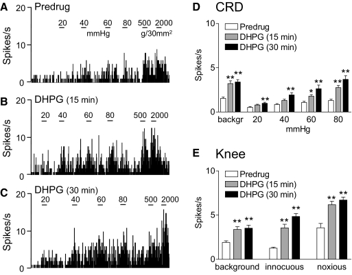
Fig. 3.
Group I metabotropic glutamate receptor (mGluR) agonist-induced facilitation. A–C: extracellular single-unit recordings of background activity and evoked responses of a CeLC neuron before drug application (predrug, A) and during administration of (S)-3,5-dihydroxyphenylglycine (DHPG, 100 μM, concentration in microdialysis probe) into the CeLC for 15 min (B) and 30 min (C). A: the neuron showed graded responses to CRD of increasing intraluminal pressure (20, 40, 60, and 80 mmHg) and knee joint compression with innocuous (500 g/30 mm2) and noxious (2,000 g/30 mm2) intensities. B and C: DHPG increased responses to CRD and knee joint compression. The effects did not show desensitization but persisted during continued drug application. PSTHs (bin width, 1 s) show spikes/s. Horizontal lines indicate stimulus duration (15 s). D and E: summary data. Bar histograms (mean ± SE) show background activity and responses evoked by CRD (D) and by knee joint compression (E) averaged across the sample of neurons (n = 6). *P < 0.05, **P < 0.01 (compared with predrug control; Dunnett's multiple comparison test).

