Fig. 5.
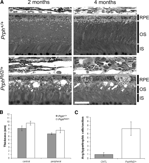
PrphRd2/+ mice develop RPE abnormalities by 4 mo. A: representative photomicrographs of 2 and 4 mo old Prph+/+ and PrphRd2/+ retina/RPE sections. Layers present are identified. RPE, retinal pigment epithelium; OS, outer segment layer; IS, inner segment layer. Images were taken with a ×100 objective; scale bar = 20 μm. Arrows indicate swollen, hypertrophic RPE cells and dimpled arrowheads specify vacuoles. B: thickness of the RPE cell layer was measured in both central and peripheral regions of the retina at 4 mo. C: the total number of hypertrophic RPE cells per section at 4 mo was counted.

