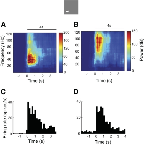
Fig. 6.
Two examples of neural responses to visual stimulation in area 17. Neural activity was recorded by the dual-purpose microsensor. Electrode penetrations were made along the medial bank of the postlateral gyrus from Horsley–Clarke coordinates P4L2 at an angle of 10° medial and 20° anterior. Sinusoidal drifting gratings at preferred orientation, spatial frequency, temporal frequency, and size were presented to the dominant eye while the other eye viewed a blank screen. Stimulus contrast was 50%. Horizontal lines represent stimulus onset and duration. (A, B) LFP spectrograms and (C, D) MUAs for 2 example recording sites.

