Fig. 1.
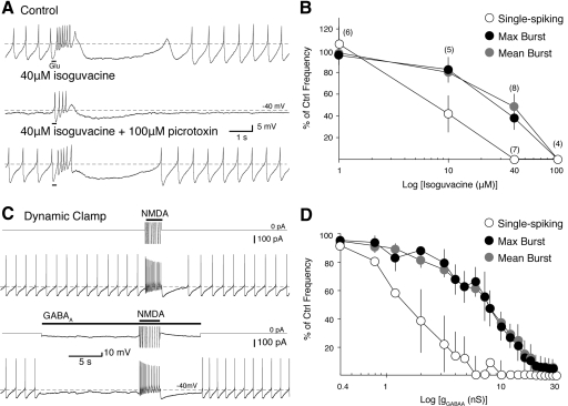
Single-spiking is more sensitive to suppression by GABAA receptor (GABAAR) activation than N-methyl-d-aspartate receptor (NMDAR)-mediated burst firing. A: a phasic burst (15 Hz maximum burst frequency) was evoked in a perforated patch recording of an identified dopaminergic neuron by iontophoresis of glutamate (Glu; black bars beneath trace, top) in 25 μM NBQX. The GABAAR agonist isoguvacine (40 μM) was applied to the bath (middle). Single-spike firing was completely suppressed while the burst frequency was decreased by 62%. The effect of isoguvacine was reversed by subsequent application of the chloride channel blocker, picrotoxin (100 μM, bottom). B: summarized data show the percent inhibition of maximum burst frequency (filled circles), mean burst frequency (gray circles), and mean spontaneous firing frequency (empty circles) from control frequencies after bath application of isoguvacine (1, 10, 40, and 100 μM). Numbers in parentheses indicate n for each concentration. C: application of a 40 nS NMDAR conductance using dynamic clamp elicits a burst (20 Hz maximum) in a whole cell somatic recording of an identified dopaminergic neuron (top). Application of a 2.8 nS GABAAR conductance (EGABAA = −60 mV) suppresses single-spike activity while the burst remains (18 Hz maximum; bottom). Above each voltage trace is the truncated current applied from the dynamic clamp (downward represents an outward current). D: summarized data (n = 4) show the percent of inhibition of maximum burst frequency (filled circles), mean burst frequency (gray circles), and mean spontaneous firing frequency (empty circles) to application of a series of GABAAR conductances in dynamic clamp (as in C).

