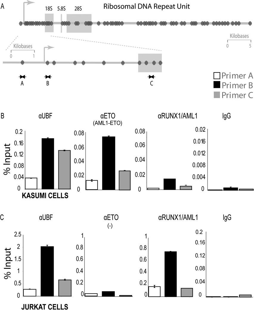
Figure 3. AML1-ETO shows enhanced occupancy of rDNA repeats during mitosis.
A) The panel shows a schematic of the Runx consensus elements (dark ovals) in the human rDNA repeats and depicts the locations of ChIP primers. B and C) ChIPs were done with antibodies for RUNX1/AML1, ETO and UBF1, as well as non immune IgG in Kasumi-1 and Jurkat cells blocked in mitosis. An antibody detecting ETO was used to immunoprecipitate the AML1-ETO fusion protein, whereas an antibody recognizing the C-terminal domain of RUNX1/AML1 was used to pull down endogenous RUNX1/AML1. Quantitative PCR data are normalized to genomic DNA and denoted as percent input.

