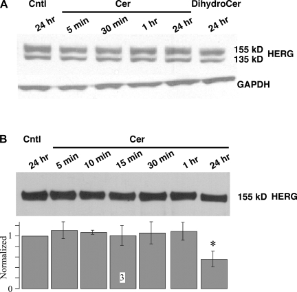
Fig. 5.
Acute Cer treatment does not affect HERG protein levels. A: HEK-293 cells were treated with either 10 μM Cer or DihydroCer for the durations indicated. Densitometric values of the mature 155-kDa band were normalized with GAPDH levels and are shown below a representative gel (n = 3). B: HEK-293 cells stably expressing HERG were treated with 10 μM Cer for the durations indicated. Cells were then treated with biotin to bind proteins expressed on the cell surface, followed by treatment with streptavidine beads. Protein bound to streptavidine beads was separated, and equal protein was loaded on the gel and probed for HERG. A representative blot is shown above, with normalized and averaged densitometer values shown in the bar graph below (n = 3). All data were normalized to the Cntl value for that run, and only the value at 24-h Cer treatment was statistically different from Cntl. *P < 0.05.

