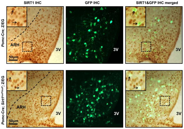
Figure 1. Deletion of SIRT1 is restricted to POMC neurons.
Representative photomicrographs of brain slices from Pomc-Cre; Z/EG (control) or Pomc-Cre; Sirt1loxP/loxP; Z/EG mice stained for SIRT1 and GFP (the Cre-conditional expression of GFP is restricted only to POMC neurons in these mice (Balthasar et al., 2004)). Dark-brown staining and green fluorescence represent SIRT1 and GFP immunoreactivity, respectively. Higher magnification of the boxed-region is in the top-left corner of the photomicrograph. Arrows indicate POMC neurons. Note the co-localization between SIRT1 and POMC in Pomc-Cre; Z/EG control brain. This co-localization is absent in almost all of POMC neurons in Pomc-Cre; Sirt1loxP/loxP; Z/EG brain. Dash lines indicate ARH boundaries. Abbreviations: third ventricle (3V), hypothalamic arcuate nucleus (ARH), immunohistochemistry (IHC). Scale bar = 50 μm.

