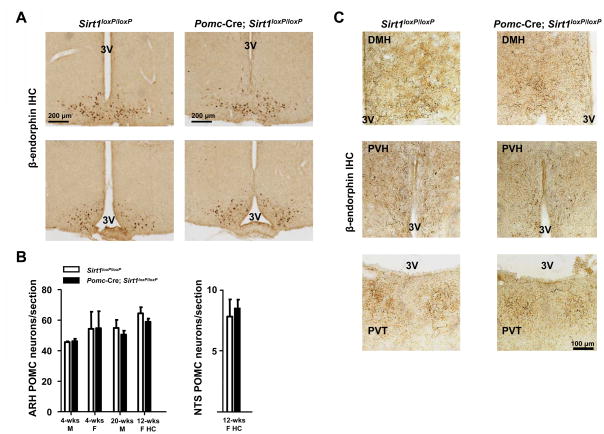
Figure 2. SIRT1 is dispensable for POMC neuronal survival.
(A) Representative photomicrographs of rostral to caudal (top to bottom) brain sections from either Sirt1loxP/loxP (control) or Pomc-Cre, Sirt1loxP/loxP mice stained for β-endorphin (a product of POMC). Dark-brown staining represents β-endorphin immunoreactivity. Scale bar = 200 μm. (B) POMC neurons were counted in several sections that contained similar regions of the hypothalamus or hindbrain in both genotypes. No difference in the estimated number of POMC neurons per section was noted between genotypes in 4-week-old males (4-wks M), 4-week-old females (4-wks F), 20-week-old males (20-wks M), and 12-week-old females fed on the high-calorie diet for 4 weeks (12-wks F HC). (C), Representative photomicrographs of brain sections from either Sirt1loxP/loxP (control) or Pomc-Cre, Sirt1loxP/loxP mice stained for β-endorphin. Dark-brown staining represents β-endorphin immunoreactive fibers. Both genotypes display similar POMC neurons fibers density in dorsomedial hypothalamic nucleus (DMH), paraventricular hypothalamic nucleus (PVH), and paraventricular thalamic nucleus (PVT). Scale bar = 100 μm. Other abbreviations: third ventricle (3V); immunohistochemistry (IHC). n=2–3 in each group. Error bars represent s.e.m. Statistical analyses were done using two-tailed unpaired Student’s t test.

