
Figure 2. Tinnitus as the result of a broken neural “noise-cancellation” mechanism.
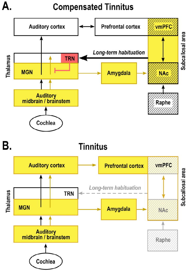
According to the theory proposed here, the nucleus accumbens (NAc) and its associated paralimbic networks in the ventromedial prefrontal cortex (vmPFC) play an important role in long-term habituation to continuous unpleasant sounds. Sound-evoked neural activity is relayed from the auditory periphery via the brainstem and thalamus (MGN) to the auditory cortex for conscious perception. The same signal is directed in parallel via the amygdala to the subcallosal area (which includes the NAc region of the ventral, “limbic” striatum and the vmPFC) for evaluation of the sound's emotional content. From the subcallosal area, an excitatory projection feeds back to the thalamic reticular nucleus (TRN), which in turn inhibits selectively the sections of the MGN corresponding to the unpleasant sound frequencies. This gain-control mechanism leads to a highly specific filtering (“tuning out”) of repetitive unwanted noises, which, as a consequence, do not reach conscious perception in the auditory cortex. The initial tinnitus signal results from peripheral deafferentation (loss of hair cells through injury, aging, or loud-noise exposure) and subsequent lesion-induced reorganization of central auditory structures. As long as the NAc-system is intact (A: “Compensated Tinnitus”), the tinnitus signal is filtered out and will not be relayed to the auditory cortex. If, however, the NAc-system becomes compromised (B: “Tinnitus”), cancellation of the tinnitus signal at the thalamic level is no longer possible, tinnitus perception results, and long-term reorganization of auditory cortex sets in to render the tinnitus chronic. Cases of intermittent tinnitus may occur during a stage at which damage to the subcallosal area is still temporary: fluctuating activity (and corresponding neurotransmitter) levels allow transient filtering of the tinnitus signal. The raphe nuclei, which control serotonin levels and sleep cycles, provide a major input to the NAc and may thus contribute to the correlation between tinnitus strength and insomnia. Structures with serotonergic innervation are shown by hatching; inhibitory structures are shown in red; yellow indicates presence of tinnitus signal.