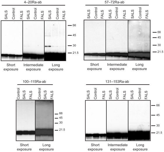
Figure 3. Analysis of SOD1 by western immunoblot.
Homogenates of tissue from the temporal lobe, the precentral gyrus, and the spinal cord ventral horns from 5 control patients, 5 SALS and 4 FALS patients were analyzed by western immunoblots, using the 4–20Ra-ab, 57–72Ra-ab, 100–115Ra-ab and 131–153Ra-ab anti-SOD1 peptide antibodies. Analyses of lumbar spinal cord ventral horns are here shown as examples. All figures depict the same set of extracts and short, intermediate and long exposures are presented. Upon long exposure a weak band at about 28 kDa was seen with the 4–20Ra-ab anti-SOD1 antibody in one of the five SALS samples. The band was probably unspecific since it was not seen with the other antibodies. The intensity was estimated at <0.5% of the SOD1 band.

