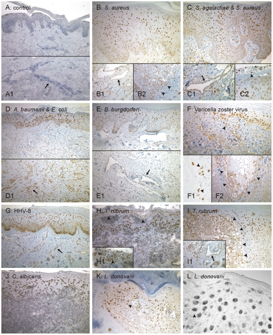
Figure 1. Detection of HIF-1 activation in human skin biopsies of patients suffering from cutaneous infections by immunohistochemistry.
(A) In uninfected skin, rare and faint nuclear HIF-1α staining occurred in the epidermis whereas the dermis was negative for HIF-1α (A1). (B to L) In all cutaneous infections, a strong nuclear HIF-1α signal was detected in keratinocytes, predominantly of the spinal cell layer (examplified in L). Moreover, endothelial cells of dermal capillaries frequently stained positive (arrows in B1, C1, D1, E1, G, I1). Arrowheads: (B2) border zone of the positive abscess; (C2): positive neutrophils; (F): positive dermal lympho-histiocytic infiltrates; (F1): large positive nuclei of a multi-nuclear giant cell; (F2): skin lesion surrounded by a HIF-1 positive inflammatory infiltrate; (H): positive sub-corneal neutrophils (higher power in H1); (I): positive intra-epidermal infiltrate; (K): intradermal nodules containing leishmaniae. (A) Control: uninfected sample; (B–E): bioptic samples of patients suffering from infections with humanpathogenic bacteria [(B) S. aureus, (C) S. agalactiae and S. aureus, (D) A. baumanii and E. coli and (E) B. burgdorferi], (F, G) with humanpathogenic Herpes-viruses [(F) Varicella zoster virus (VZV) and (G) Human Herpes Virus-8 (HHV 8)], (H–J) with humanpathogenic fungi [(H+I) T. rubrum and (J) C. albicans] or (K, L) with the protozoic pathogen L. donovani. Magnifications: all 250×, except for A1, B1, B2, C1, D1, E1, F2, I1 (all 400×), and C2, F1, H1, L (all 1,000×).

