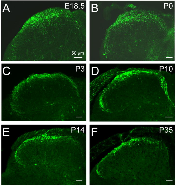
Fig. 3.
Distribution of the central projections of TRPM8 fibers in the developing spinal dorsal horn. At E18.5 and P0, Trpm8GFP axons are densely innervating the dorsal horn, but largely restricted to the medial sites (A, B). By P3 and P10, Trpm8GFP axons have reached lateral regions of the dorsal horn (C, D) and by P14 (E) have resolved into an adult-like somatotopic organizational pattern (F). Bars equal 50 μm.

