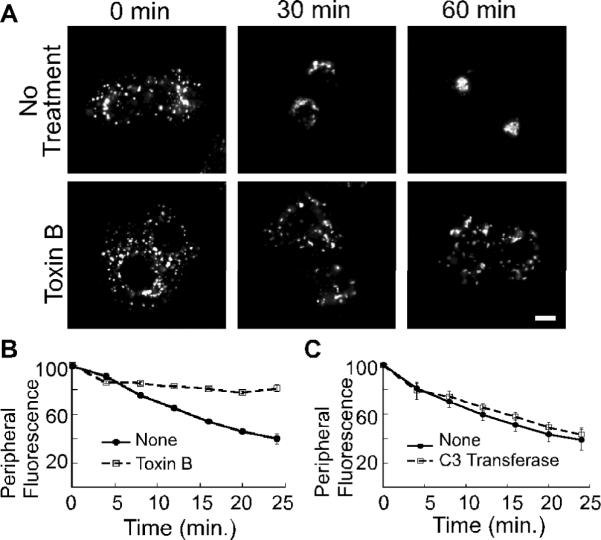
Figure 2. Microtubule-dependent Golgi positioning requires Rho GTPases.

(A) NRK cells were treated with nocodazole for 2 hours. 100 ng/ml Toxin B was added as the nocodazole was washed out. Shown are confocal micrographs of cells that were fixed at the indicated time points and decorated with anti-GM130 antibody. The size bars represent 5 μm. (B–C) Golgi motility in living NRK cells was quantified by incubation with NBD-C6 ceramide prior to nocodazole addition. 100 ng/ml Toxin B (B) or 2 μg/ml C3 transferase (C) was added during the nocodazole wash out. A peripheral region of interest was defined for each cell and the change in peripheral fluorescence was recorded for 25 minutes at 37°C. The mean fluorescence intensity within the region of interest is plotted as a function of time; n=3 experiments for (B,C). The standard error in each case is indicated by bars.
