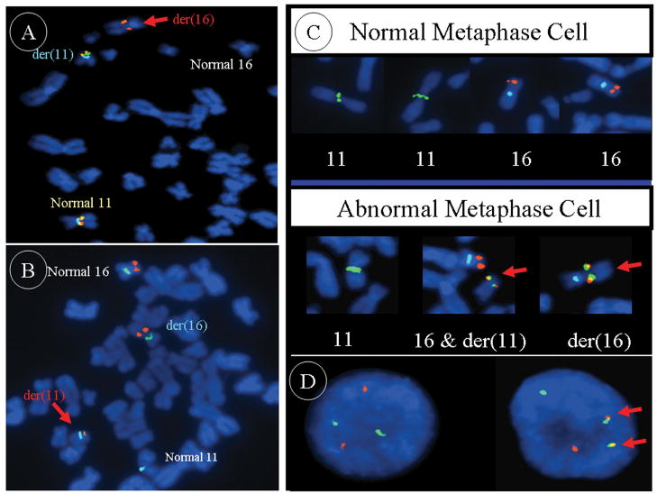
Figure 3.
A: The 11q13 breakpoint is shown to lie within the spectrum orange labeled BAC probe RP11-466C23 (BAC probe CTD-3110P2 is labeled in green and CEP11 in aqua). B: The 16p13 breakpoint is shown to lie within the spectrum orange labeled BAC probe CTD-2118O12 (CEP16 is labeled in green and CEP11 in aqua). C: The dual-color dual-fusion probe set (RP11-697H9 and RP11-466C23 cocktail probe labeled in spectrum green, CTD-2135D7 and CTD-2118O12 cocktail probe in orange, and CEP16 in aqua) confirms the presence of the 11;16 translocation on each of the respective derivative chromosomes in case 1 (lower panel) in contrast to single signals on the chromosome 11 and 16 homologues from a normal peripheral blood lymphocyte metaphase cell (upper panel). D: The dual-color dual-fusion probe set in a normal interphase cell (left) and in a translocation positive chondroid lipoma interphase cell (right).

