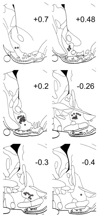
Figure 1.
Histological localization of potassium ferrocyanide lesioned microwire tips in serial coronal sections (Paxinos and Watson, 1997). Number on each plate indicates anteroposterior distance (mm) from bregma. Each filled circle represents a single microwire within the VP that recorded neural activity.

