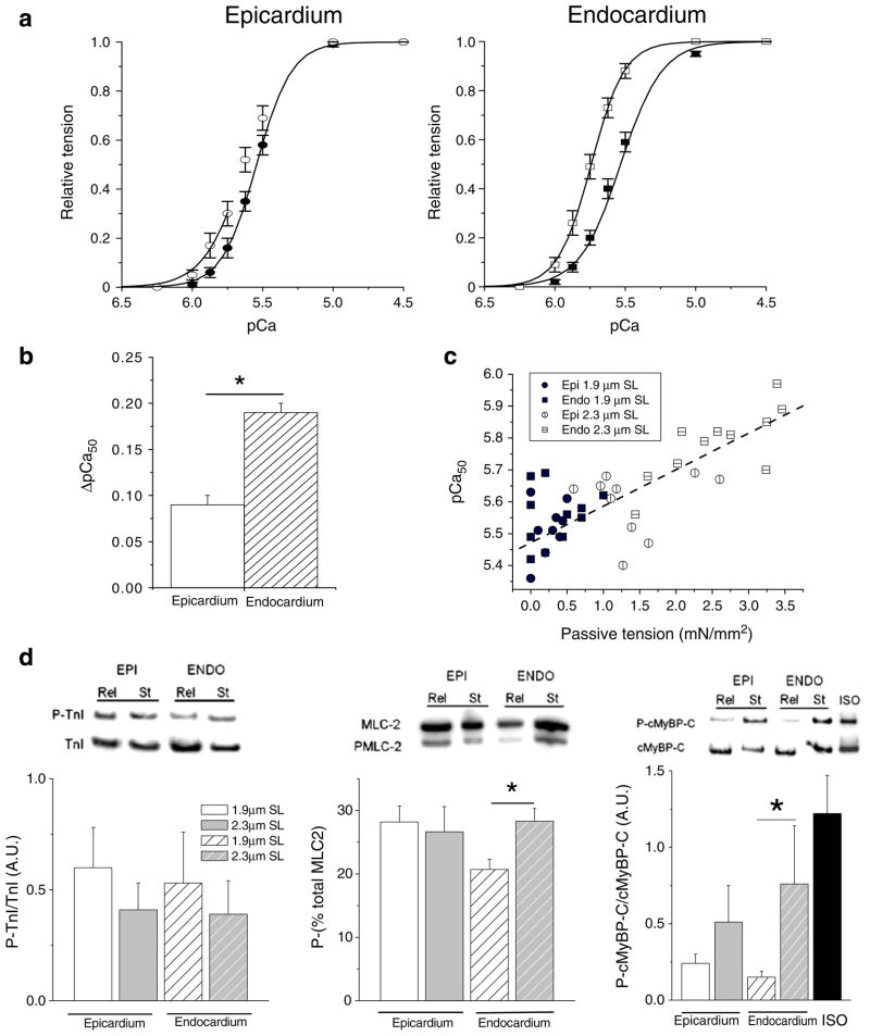
Fig. 2.
Relationship between the relative tension and calcium in ENDO and EPI cells at two sarcomere lengths. a Changes of calcium sensitivity of the contractile machinery are indicated by the leftward shift of the tension–pCa curve when cells were stretched from 1.9 μm SL (closed symbols) to 2.3 μm SL (open symbols; n=14 cells per region). b Stretch-induced calcium sensitization (ΔpCa50) was more prominent in ENDO cells. c Relationship between calcium sensitivity and passive tension for all data from ENDO and EPI at two sarcomere lengths. Passive tension was measured in relaxing solution prior each activation procedure from slack length to 1.9 or 2.3 μm SL (y=5.47+ 0.011x, r=−0.82, P<0.001). d Skinned muscle strips dissected from the sub-epicardial layer (full bar) or the sub-endocardial layer (hatched bar) were quick-frozen either at slack length (open bar) or after stretch to 20–30% of the slack length (grey bar). Left: immunoblots with anti-cardiac TnI and anti-cTnI phosphorylated by PKA showed that TnI phosphorylation was not affected by stretch (n= 6 animals/group). Middle: for MLC-2 phosphorylation quantification, each isoform was separated and expressed as a percentage of the total amount (phosphorylated + non-phosphorylated forms). Right: Immunoblots with anti-cardiac MyBP-C and anti-phospho Ser282 cMyBP-C showed that cMyBP-C phosphorylation level was increased by stretch (n=6 animals/group). Isoprenaline stimulation (Iso) of intact myocytes was used as control for full phosphorylation of MyBP-C. *P<0.05

