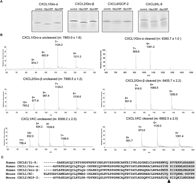
Fig. 1.

SpyCEP cleaves human CXCL1, 2, 6 and 8 and murine CXCL1/2 after the first four amino acids of the C-terminal α-helix. A. Colloidal Coomassie blue stained SDS-PAGE gels showing human chemokines cleaved by SpyCEP. Chemokines were co-incubated either alone (left lane); with supernatant containing SpyCEP, H292 (+SpyCEP, middle lane); or supernatant without SpyCEP, H575 (-SpyCEP, right lane). B. Mass spectroscopy analysis of uncleaved and cleaved chemokines using electrospray ionization generated a series of multiply charged ions (indicated as m/z; mass-to-charge ratio) from which the average molecular mass (m) of each was deduced. C. Site of SpyCEP-mediated cleavage of various chemokines determined in this study and previously for CXCL8/IL-8 and CXCL2/MIP-2 (Edwards et al., 2005): arrowhead. The position of the α-helix in this region is indicated by a grey shaded box.
