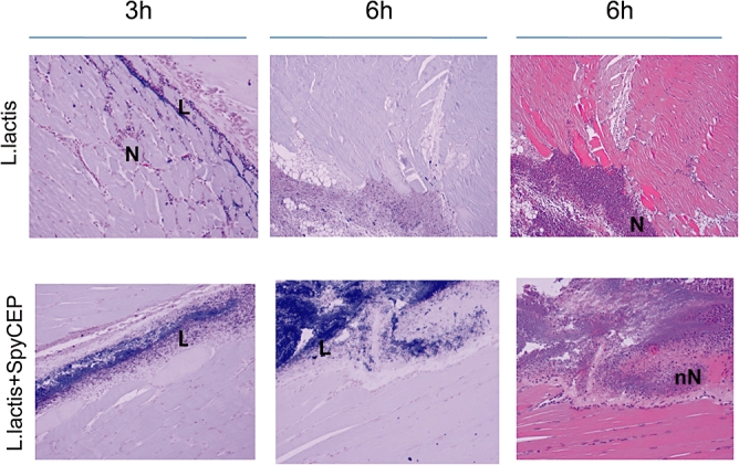
Fig. 5.

Tissue sections from thigh muscle of L. lactis-infected mice obtained at 3 and 6 h after onset of infection suggest that SpyCEP is sufficient to reproduce some features of invasive necrotizing infection. Gram stained muscle sections from L. lactis-infected mice at 3 and 6 h after infection (left and central panels). Upper panels, mice infected with control L. lactis H486 (empty plasmid); Lower panels, mice infected with L. lactis strain expressing SpyCEP, H487. Right hand panels, haematoxylin and eosin-stained tissues at 6 h. L, lactococci; N, neutrophils; nN, necrotic neutrophil. Representative of three mice at each time point, magnification ×200.
