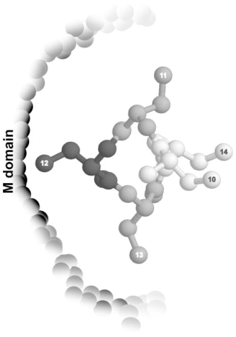
FIGURE 3.
Schematic representation of the hydrophobic core of the signal peptide (seen from the top) bound to the M domain of Ffh in a helical conformation, as suggested by the positional dependence of BPM-mediated crosslinking. The intensity of the gray shading for different residues indicates the crosslinking efficiency at that position.

