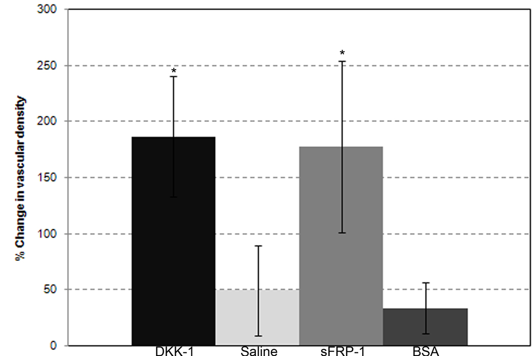
Figure 3.
Changes in vascular density. Each column represents the mean change +/− SD over three days of vascular density in the mesentery windows of the DKK-1 (n=7), sFRP-1 (n=5), saline (n=6) and BSA (n=4) treatments, respectively. The vascular density measurement was described in the Materials and Methods. * - Significant compared to both saline and BSA with p < .05.

