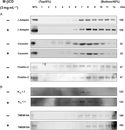
Figure 6.
Effect of M-βCD on lipid raft-enriched fractions prepared from murine portal vein. (A) Western blot analysis of membrane proteins separated by a discontinuous sucrose density gradient (0, 20, 25, 30, 35, 40% sucrose) in the absence (−) and presence of 3 mg mL−1 M-βCD for 15 min (+). (B) A representative western blot for fractionated proteins probed with antibodies against KCa1.1 and TMEM16A ± M-βCD for 15 min.

