Figure 5.
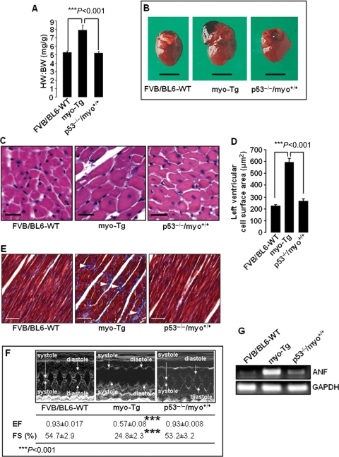
Absence of p53 in p53−/−/myo+/+ double Tg mice resulted in the regression of cardiac mass associated with improved cardiac function. (A) HW:BW ratios showing significant (Bonferroni's corrected P < 0.001) reduction in cardiac mass in p53−/−/myo+/+ mice compared with myo-Tg mice. The results are presented as mean ± SEM and represent three independent experiments. (B) Hearts from 16-week-old FVB/BL6-WT, myo-Tg, and p53−/−/myo+/+ mice; (C) Haematoxylin–eosin stained cells from left ventricles of FVB/BL6-WT, myo-Tg, and p53−/−/myo+/+ mice (×20 magnification, white bar represents 25 µm); (D) Measurement of left ventricular cell surface area showing significantly enlarged cell (Bonferroni's corrected P < 0.001) in myo-Tg mice compared with FVB/BL6-WT and p53−/−/myo+/+ mice. Data (mean ± SEM) for each group is an average of nine individual measurements; (E) Masson's trichrome staining of the left ventricle demonstrates collagen deposition (white arrowhead) in myo-Tg mice (×20 magnification, white bar represents 50 µm); (F) M-mode echocardiography showing improved cardiac function in p53−/−/myo+/+ mice, where values for both ejection fraction (EF) and fractional shortening (FS) increased significantly (Bonferroni's corrected P< 0.001) compared with myo-Tg mice and are almost identical to those of WT mice; (G) RT-PCR analysis showing downregulation of the hypertrophic marker gene atrial natriuretic factor (ANF) in p53−/−/myo+/+ mice. In RT-PCR analysis, GAPDH was analysed as a loading control.

