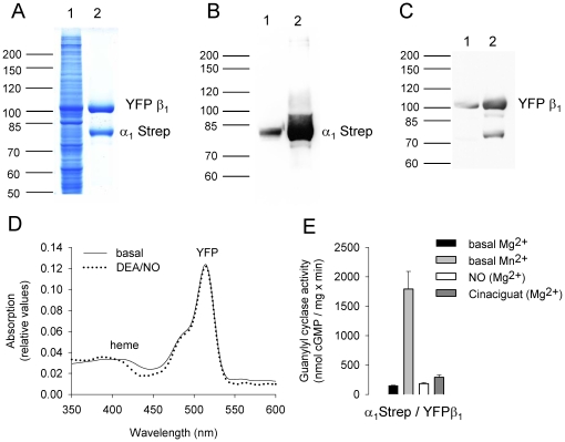
Figure 2. Analysis of purified heterodimeric sGC with an amino-terminally tagged β1 subunit.
Coomassie blue staining (A) and Western blot analysis with antibodies directed against the α1 subunit (B) and the β1 subunit (C) using cytosolic fraction (lane 1) or purified sGC (lane 2). Absorption spectra of purified sGC under basal conditions (solid line) and in the presence of 100 µM DEA/NO (dotted line) (D). Specific sGC activity of purified sGC assayed with Mg2+ (black column) and Mn2+ (grey column) as cofactor and in the presence of 100 µM DEA/NO (white column) and 10 µM Cinaciguat (dark grey column) (E).

