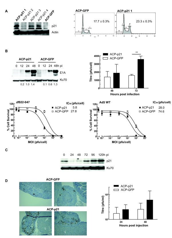
Figure 5.
p21 re-expression increases adenovirus activity. 5A: Expression of p21 in A2780CP cells increases S phase fraction. A2780CP expressing p21 cells were generated as described in Materials and Methods. Expression of p21 in one pool (ACP-p21-1) was confirmed by immunoblot (left). Cell cycle status in asynchronous ACP-p21-1 and control cells (ACP-GFP) was assessed following propidium iodide staining. Figures represent the percentage cells in S phase. 5B: p21 expression increases E1A expression, viral replication and cytotoxicity. 105 ACP-p21 and ACP-GFP were infected with dl922-947 (MOI 10). E1A expression was assessed by immunoblot (top left) and intracellular virion production was assessed by TCID50 (top right) ** p < 0.01. ACP-p21 and ACP-GFP cells were infected with dl922-947 and Ad5 WT (MOI 0.01-1000). Numbers below blots represent E1A:Ku70 ratio. Cell survival was assessed 120 hours later (5B bottom left and right). 5C: p21 expression in ACP-p21 cells decreases following dl922-947 infection. ACP-p21 cells were infected with dl922-947 (MOI 10). Expression of p21 was analyzed by immunoblot up to 120 h pi. 5D: p21 expression increases viral activity in vivo. Female Balb C nu/nu mice were inoculated ip with 5 × 10⁶ ACP-p21 or ACP-GFP cells (n = 5 per group). One week later, dl922-947 was injected ip (5 × 10⁹ particles daily x3). Blood was taken 24 hours after last virus injection and mice were killed 24 h thereafter. Expression of E1A was assessed by immunohistochemistry (left). All images are x100, S = Small intestine; L = Liver. Virion levels in serum were assessed by TCID50 (right). Results represent mean ± sd, n = 5.

