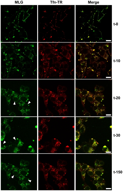
Figure 3. Uncoating and distribution of M protein in live cells.
Cells were inoculated with rVSV-MLG and Tfn-TR at 4°C for 90 minutes and either fixed immediately with ice-cold 3% paraformaldehyde (t-0) or were quickly warmed by the addition of 37°C media and examined without fixation by LSCM on a heated stage at the indicated times using separate 35mm glass bottomed dishes for each time point to reduce signal loss by photobleaching. Bars = 10 µm. Arrowheads indicate MLG localization to NPCs.

