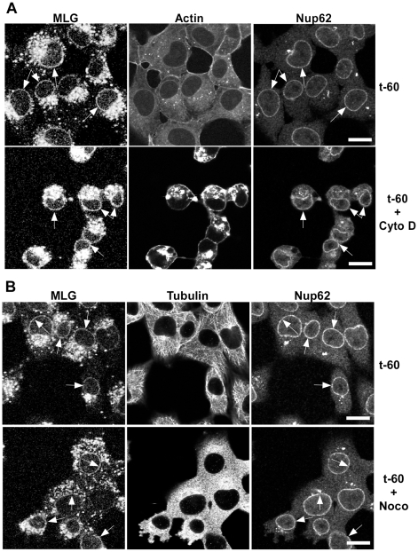
Figure 5. Distribution of MLG in the presence of cytochalasin D or nocodazole.
BHK cells were infected with rVSV-MLG in the presence of NH4Cl as described for Fig. 4B. Cytoskeletal inhibitors were added for 30 minutes before NH4Cl washout and the inhibitors remained in the post-washout media until the cells were fixed 60 minutes later. Distribution of MLG, Nup62 and actin (A) or tubulin (B) either without (top panels) or with (bottom panels) cytoskeletal inhibitors. Arrows indicate nuclear envelope localization. The brightness levels were adjusted for all the images using Canvas 11 software to maximize the MLG signal after conversion to grayscale. Bars = 10 µm.

