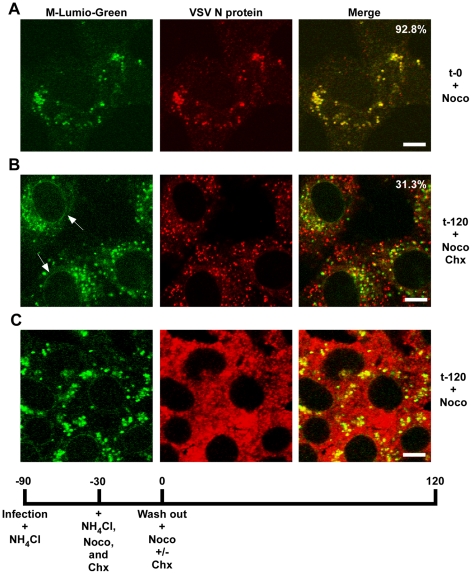
Figure 9. Release of RNPs from endosomes and viral protein synthesis in the presence of nocodazole.
Confocal images of cells inoculated with rVSV-MLG at t-0 (A) and t-120 (B and C) post-NH4Cl washout in the presence of cycloheximide and nocodazole (B), or just nocodazole (C), and stained for VSV N protein using N mAb conjugated to Alexa Fluor-568. Quantification of the amount of colocalization was determined for 50 individual cells using the colocalization function in the LSM software version 3.2. Percent of N protein that colocalized with MLG is indicated for t-0 and t-120 (n = 50 cells). Bars = 5 µm.

