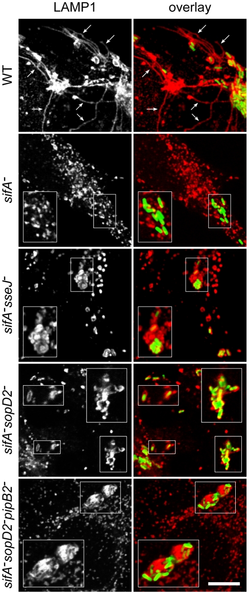
Figure 2. Immunofluorescence imaging of vacuoles enclosing various Salmonella strains.
HeLa cells infected for 16 hours with GFP-expressing Salmonella strains were immunostained for the SCV marker LAMP1 and imaged by confocal microscopy for GFP (green) and LAMP1 (red). Insets illustrate the absence of vacuole enclosing sifA − bacteria and the shapes of sifA−sseJ−, sifA−sopD2− and sifA−sopD2−pipB2− vacuoles magnified twice. SIFs (arrows) were only seen in wild-type-infected cells. Scale bar, 10 µm or 5 µm for insets.

